BioImaging Research
NLR is building new capabilities in chemical imaging to reveal components in bioenergy-relevant organisms without specimen damage and at a higher resolution.

Visualizing and tracking dynamic, often compartmentalized, metabolic events in living plants, algae, and fungi is a perpetual challenge in biology. Real-time imaging of biochemical processes will help us understand how organisms respond to their surroundings and manage energy flows.
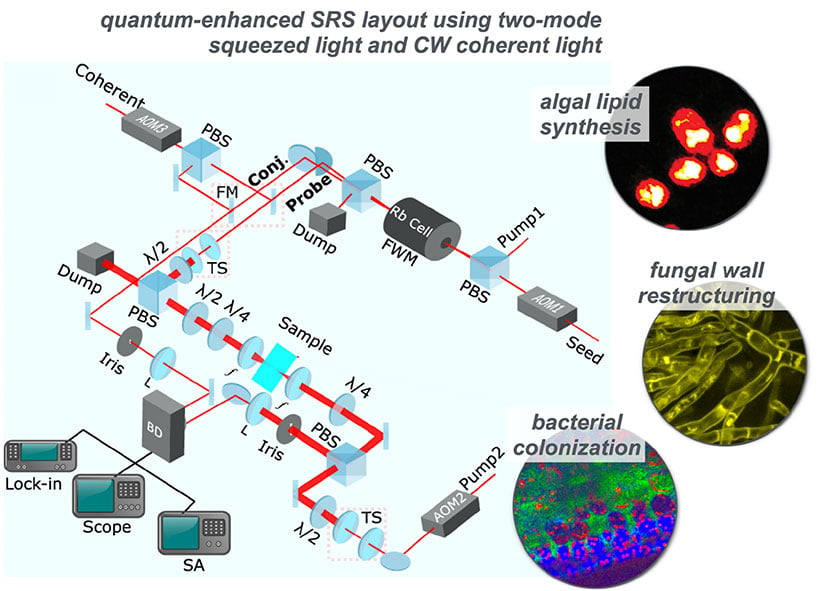
To meet these challenges, NLR is designing, assembling and integrating a prototype for a next-generation stimulated Raman scattering (SRS) microscope. The super-resolution microscope uses squeezed light and structured illumination to provide extended observation and chemical probing of biological events without jeopardizing the specimen's structural integrity or dynamics.
NLR researchers are also using quantum interference and correlations between entangled photon pairs to develop multi-modal imaging with undetected photons. This approach transfers image information from the infrared to the visible, where a standard camera can detect it.

Conceptual project workflow of novel approaches being assimilated into SRS microscopy.
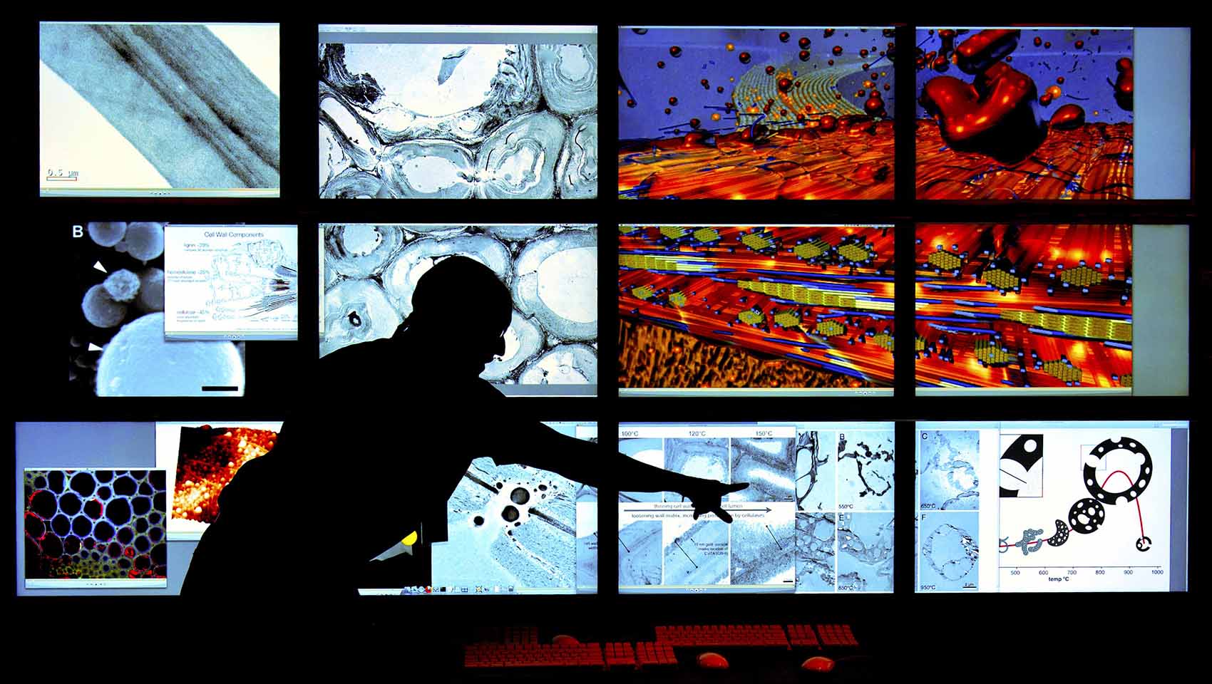
Biological Structure and Characterization Laboratory
To learn more about NLR's multi-modal imaging capabilities, read about the Biomass Surface Characterization Laboratory.
Significance and Impacts
With these innovative microscopy tools, chemical imaging investigations will be expanded, and photodamage reduced to accommodate extensive spatial and temporal dimensions.
Contact
Share
Last Updated Dec. 6, 2025
